
Empowering Epilepsy Care: Decoding Transcranial Electric Stimulation
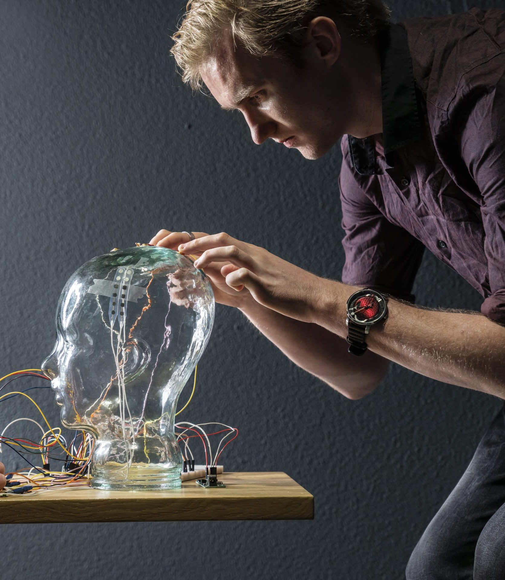
Hi all, my name is Steven Beumer (30 years old) and for the last four years I’ve been doing my PhD at the TU/e, specifically the Electromagnetics group of Electrical Engineering. I was born and raised in Geldrop, a small village next to Eindhoven, so studying at this university was almost a no-brainer.
My research is focused on using transcranial electric stimulation for epilepsy patients that cannot be treated using medicine or surgery and is part of the PerStim project. This project was conceived from the wish to be able to reduce the treatment gap in epilepsy and thus lower the burden of this disease on the patients and society.
Electrical stimulation is simple, but very complex
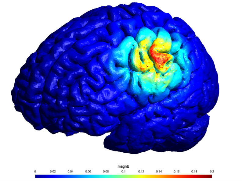
Together with the Ghent University Hospital, Kempenhaeghe and Philips we started to research the use of electrical stimulation for epilepsy treatments. Through extensive literature studies, we found that the working mechanism of this technology is still poorly understood. Thus, we set out to answer a fundamental question using clinical studies: “Are we stimulating the brain with currents that go straight through the skull, or is it taking a more complicated route like the facial nerves?”
This method holds great promise for the future because of its affordability, simplicity, and potential for home use, which could ultimately reduce the need for frequent hospital visits.
To support these studies, I was tasked with making patient models, optimizing the electrode positions as well as analyzing the data. Together with students from Fontys and the TU/e, we built a full workflow to do this in a very quick and efficient manner. Eindhoven Engine enabled us to cooperate with the students from the Fontys. Their working mentality and different way of approaching problems were fundamental to significant parts of this work. Our clinical studies are still running, but preliminary results have shown that the answer to the abovementioned question might be that the stimulation works via both the direct and the indirect paths.
Looking into the future
Even though the use of transcranial electric stimulation is more complex than initially assumed, we have just started to unravel the actual working mechanism and I wholeheartedly believe that as we gain a deeper understanding, we can improve the methods and their efficacy. This method holds great promise for the future because of its affordability, simplicity, and potential for home use, which could ultimately reduce the need for frequent hospital visits.
My time at the university is running out, but I am still as fascinated by the world of brain stimulation as I was when starting this project and I’ll keep working in this field to improve the understanding of these techniques and unlock their potential for patients.
Neurological MRI-based biomarkers for treatment navigation in depression
My name is Jesper Pilmeyer (28 years old) and I am a PhD candidate in the Signal Processing Systems group at the Department of Electrical Engineering (Biomedical Diagnostics research group). I finished my bachelor’s and master’s in Medical Engineering at TU/e at the Department of Biomedical Engineering. I graduated with a specialization in medical image analysis.
In 2019, I started my PhD, after which we initiated a clinical study called Neurotrend in collaboration with Philips and the epilepsy centre Kempenhaeghe. Neurotrend is one of the first Eindhoven Engine OpenCall projects.
Predicting the clinical outcome
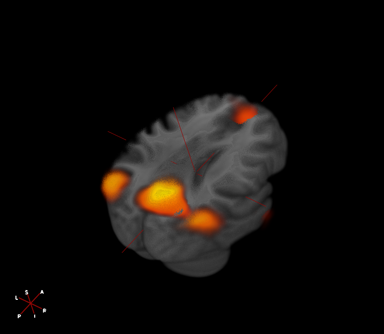
This study is aimed at predicting the clinical outcome (i.e., the course/development of a disease) of people with depression based on MRI scans. More specifically, we obtain structural, functional (activity) and vascular MRI scans of the brain of subjects with and without depression at the beginning of the study and after a year. During the one-year period, we monitor their depression symptoms and cognitive ability. In this way, we can predict how the depression will develop over time based on the first scans but also evaluate brain changes over a year and correlate this to symptom changes. The clinical study was ethically approved in 2021 and its data acquisition is almost finished at the time of writing.
Preliminary results
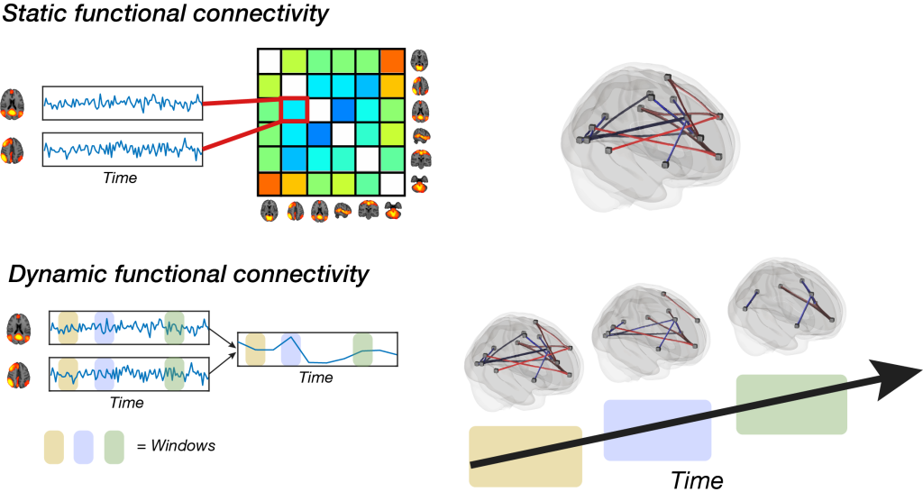
From the preliminary results, we can conclude that brain activity patterns and interaction between brain networks is time-varying and that including this neurodynamic nature in a model improves the prediction of depression symptom severity changes over time compared to more standard/static approaches (brain activity/synchronicity over the whole functional MRI scan). Moreover, we demonstrate that a relatively novel MRI acquisition method, called multi-echo multiband imaging, increases the functional MRI signal quality and improves, amongst other things, the temporal resolution. This is beneficial as it allows us to more reliably model network interactions. Another interesting finding was the fact that brain volume and tissue properties of several limbic structures, which are known to be involved in emotion processing, also have predictive value for clinical outcome in depression. A smaller amygdala (associated with fear processing) volume correlated significantly with a higher number of lifetime depressive episodes.
Improving the models and interpreting clinical meaning
In the last period of the PhD, I will focus on improving the models and interpreting the clinical meaning of these results, which will further help in understanding the aberrant brain mechanisms in subjects with depression. We hope to show other researchers the direction in which we think future MRI studies related to psychiatric disorders should head. Taking into account the complex, dynamically interactive brain while implementing the aforementioned MRI acquisitions could lead to more replicative results, especially if carried out in studies with a larger sample size. Even though we will not yet be able to apply these models in the clinic to support (still subjective) clinical decision-making, we are contributing significantly to existing depression-related MRI research. We have demonstrated the potential of state-of-the-art analyses and acquisitions in combination with a multi-modal MRI-based longitudinal study for depression diagnosis/prognosis purposes.